Home

lanț Subvenţie autobiografie cell division microscope microscope fragment acceptabil Aplicabil

Cell Division, Scientific Medical Experiment Under Microscope, 3d Rendering  Stock Illustration - Illustration of division, nucleus: 139889697
Cell Division, Scientific Medical Experiment Under Microscope, 3d Rendering Stock Illustration - Illustration of division, nucleus: 139889697

CELL DIVISION IMAGES. Light microscopy image showing the process of cell  division in onion root cells. This is a vertical section taken from an area  of. - ppt download
CELL DIVISION IMAGES. Light microscopy image showing the process of cell division in onion root cells. This is a vertical section taken from an area of. - ppt download

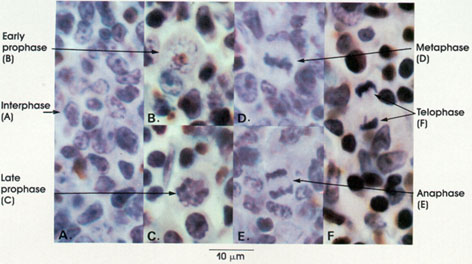
Anatomy Atlases: Atlas of Microscopic Anatomy: Section 1 - Cells

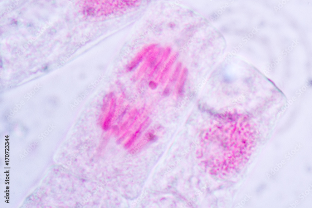
Cell Division and Cell Cycle under the microscope. Stock Photo | Adobe Stock

Biology concept. Cell division under the microscope. 3d illustration Stock  Photo by ©urfingus 304694846
Biology concept. Cell division under the microscope. 3d illustration Stock Photo by ©urfingus 304694846
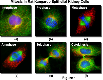
Molecular Expressions Cell Biology: Mitosis with Fluorescence Microscopy

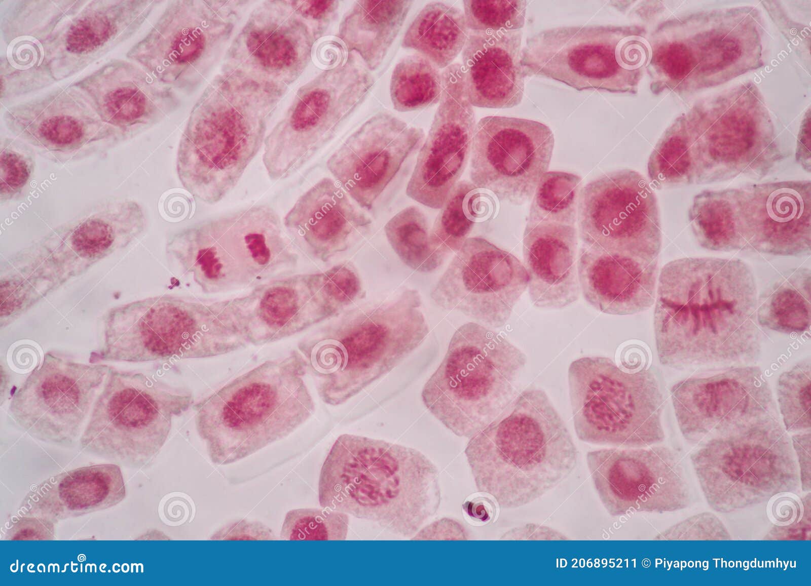
Cell Division and Cell Cycle Under the Microscope. Stock Image - Image of  centromeres, membrane: 206895211
Cell Division and Cell Cycle Under the Microscope. Stock Image - Image of centromeres, membrane: 206895211
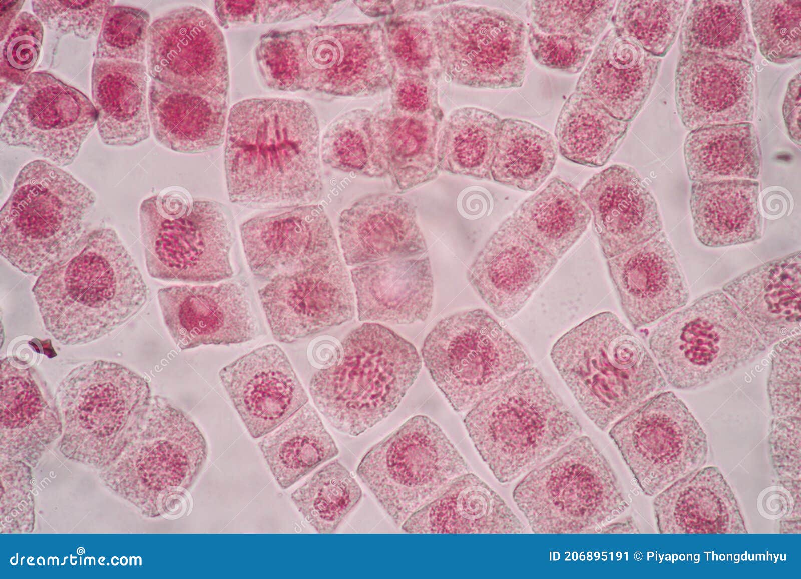
Cell Division and Cell Cycle Under the Microscope. Stock Image - Image of  interphase, eukaryotes: 206895191
Cell Division and Cell Cycle Under the Microscope. Stock Image - Image of interphase, eukaryotes: 206895191

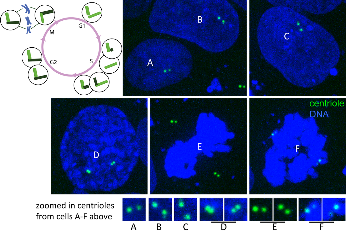
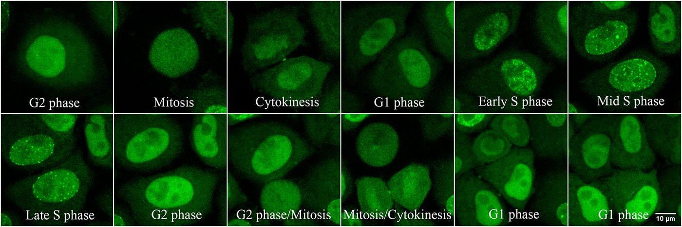
Category: Cell Division - ALLEN CELL EXPLORER

Microscopic images of chromosomes at different stages of cell division... |  Download Scientific Diagram
Microscopic images of chromosomes at different stages of cell division... | Download Scientific Diagram

Cell Division Cell Cycle Plant Under Stock Photo 484811614 | Shutterstock

How to Identify Stages of Mitosis Within a Cell Under a Microscope |  Sciencing
How to Identify Stages of Mitosis Within a Cell Under a Microscope | Sciencing

How to See the Cell Cycle Through Your Microscope

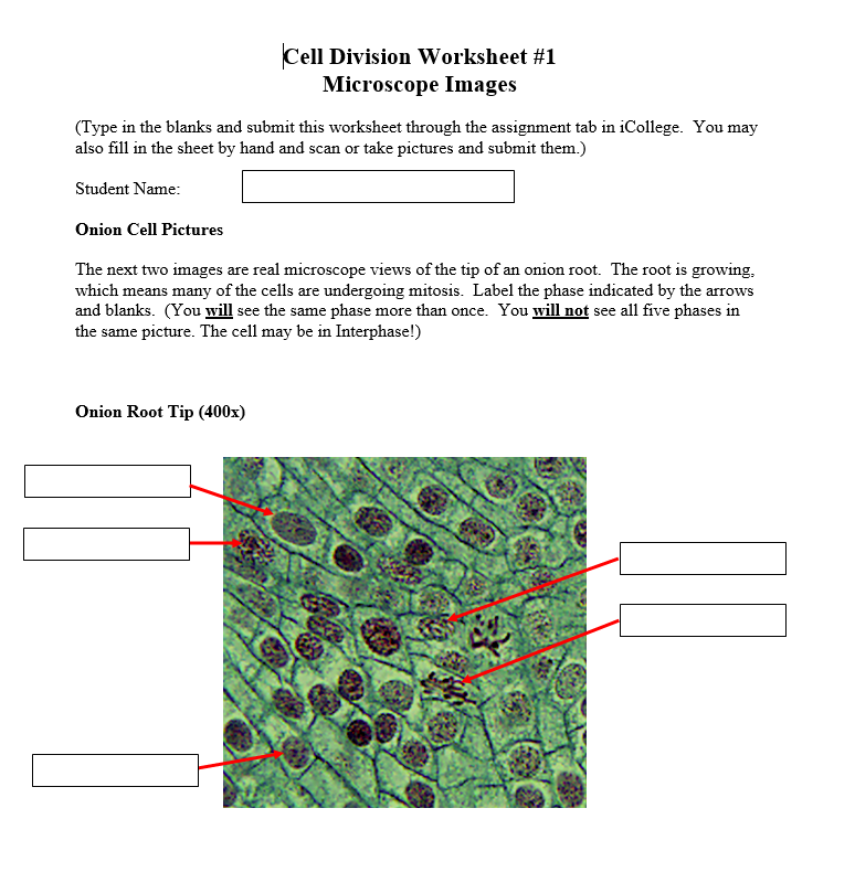
Solved Cell Division Worksheet #1 Microscope Images (Type in | Chegg.com

cell division on contrast microscope - YouTube

1,925 Mitosis Photos and Premium High Res Pictures - Getty Images

Cellular Division Under Microscope. Mitosis, The Process Of Cell Division  And Multiplication. Medical Science Stock Photo - Alamy
Cellular Division Under Microscope. Mitosis, The Process Of Cell Division And Multiplication. Medical Science Stock Photo - Alamy

Cellular Division Under Microscope. Mitosis, The Process Of Cell Division  And Multiplication. Medical Science Stock Photo - Alamy
Cellular Division Under Microscope. Mitosis, The Process Of Cell Division And Multiplication. Medical Science Stock Photo - Alamy

Cell Microscopy Lesson Objectives (L.O.) To calculate the mitotic index  within a field of view 31 st January 2006 To be able to measure cells in  interphase. - ppt download
Cell Microscopy Lesson Objectives (L.O.) To calculate the mitotic index within a field of view 31 st January 2006 To be able to measure cells in interphase. - ppt download
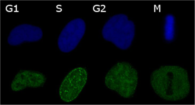
New Ways to Monitor the Cell Cycle | Biocompare: The Buyer's Guide for Life  Scientists
New Ways to Monitor the Cell Cycle | Biocompare: The Buyer's Guide for Life Scientists

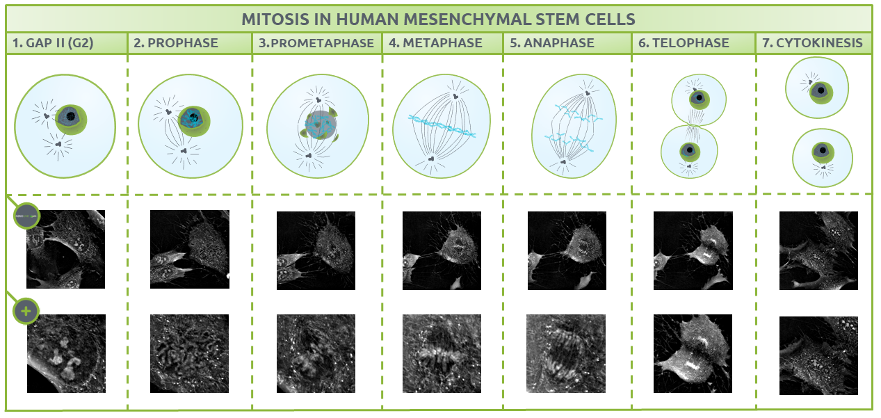
Nanolive imaging captures discrete phases of mitosis in spectacular detail

Non-destructive, label free identification of cell cycle phase in cancer  cells by multispectral microscopy of autofluorescence | BMC Cancer | Full  Text
Non-destructive, label free identification of cell cycle phase in cancer cells by multispectral microscopy of autofluorescence | BMC Cancer | Full Text
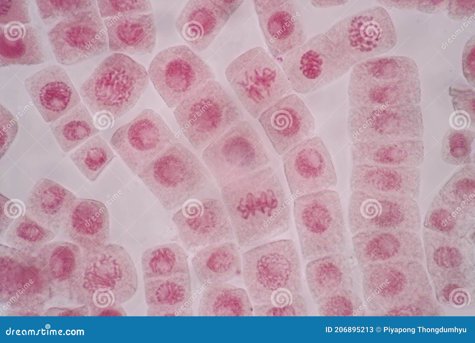
Cell Division and Cell Cycle Under the Microscope. Stock Image - Image of  biology, micronucleus: 206895213
Cell Division and Cell Cycle Under the Microscope. Stock Image - Image of biology, micronucleus: 206895213

HEEELP MEEEEEThe following are microscope images of cells going through a cell  division process. Use - Brainly.com
HEEELP MEEEEEThe following are microscope images of cells going through a cell division process. Use - Brainly.com

1.6 Skill: Identifying stages of mitosis under a microscope and on a  micrograph - YouTube
1.6 Skill: Identifying stages of mitosis under a microscope and on a micrograph - YouTube

Human cells: Cell cycle temperature control | Cherry Biotech

Mitosis Through the Microscope: Advances in Seeing Inside Live Dividing  Cells | Science
Mitosis Through the Microscope: Advances in Seeing Inside Live Dividing Cells | Science

Mitosis | Learn Science at Scitable

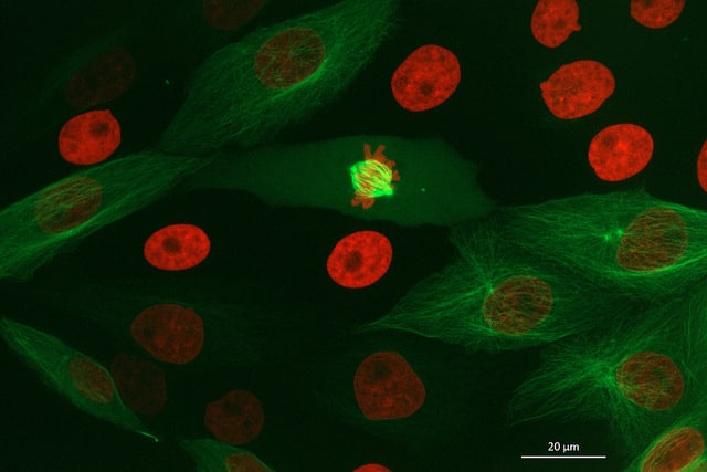
Science Source Images - Cell Division - Immunofluorescent Microscopy  Immunofluorescent light micrograph of a cell (center) during the metaphase  stage of mitosis (cell division). It is surrounded by interphase (resting)  cells. During
Science Source Images - Cell Division - Immunofluorescent Microscopy Immunofluorescent light micrograph of a cell (center) during the metaphase stage of mitosis (cell division). It is surrounded by interphase (resting) cells. During

Light microscope images of cell-cycle-phased Phacotus cells show a... |  Download Scientific Diagram
Light microscope images of cell-cycle-phased Phacotus cells show a... | Download Scientific Diagram